Left): Porcine ventricle sample, epicardium side up, mounted to
Download scientific diagram | (Left): Porcine ventricle sample, epicardium side up, mounted to the silicone lined fixture with Tpins. (Right): Porcine aorta sample, intima side up, mounted to the silicone lined fixture with T-pins. (Both): 0.25 in diameter steel ball upper member as test probe. from publication: PolyJet 3D Printing of Tissue Mimicking Materials: An Investigation of Characteristic Properties of 3D Printed Synthetic Tissue | Current anatomical 3D printing has been primarily used for education, training, and surgical planning purposes. This is largely due to the models being printed in materials which excel at replicating macro-level organic geometries; however, these materials have the drawback | 3D Printing, Tissue and Subcutaneous Tissue | ResearchGate, the professional network for scientists.
The mesh of the left and right ventricle, showing epicardium and

PDF) PolyJet 3D Printing of Tissue Mimicking Materials: An

Transverse Shear Along Myocardial Cleavage Planes Provides a

JCDD, Free Full-Text
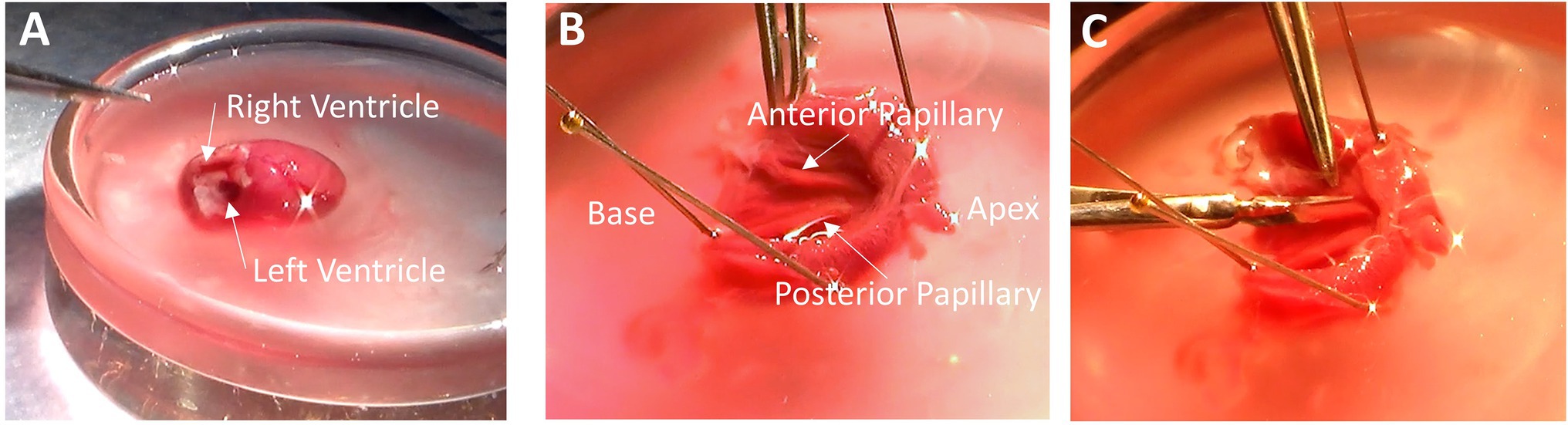
Frontiers Preparing Excitable Cardiac Papillary Muscle and
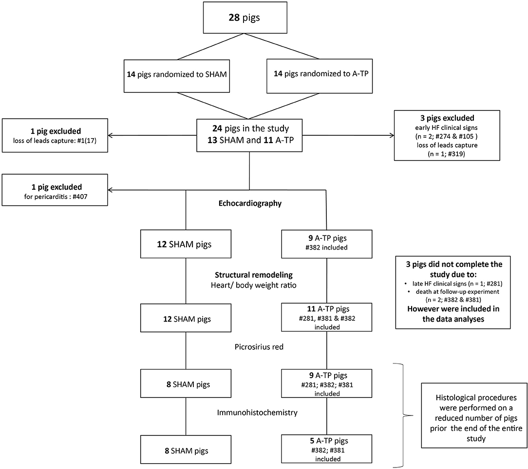
Frontiers Characterization of Atrial and Ventricular Structural

A 3D high resolution MRI method for the visualization of cardiac

Heart - Wikipedia

Close-up of the lateral view of the left side of the heart with

Medicina, Free Full-Text

Morphology, distribution, and variability of the epicardiac neural

PDF) PolyJet 3D Printing of Tissue Mimicking Materials: An

E9.5-E10.5. The proepicardial organ and the first covering of the

Reversal of Right Ventricular Remodeling After Correction of

Biventricular biaxial mechanical testing and constitutive